Radiographs (x-ray) in dentistry are now the standard. They allow eg to detect caries that begins between the teeth or become familiar with the anatomy of canals and roots in case of root canal treatment. X-ray of the tooth allows the doctor to diagnose better and more accurately, and hence, treat precisely.
Types of X-ray pictures
Depending on the case, you may need to take a specific type of X-ray. There are:
Cephalometric X-ray – this is a side view of the skull on which we see soft tissues and hard as well as paranasal sinuses and hard palate. This type of picture enables detection of malocclusion and planning of optimal treatment in a given case, it is very often used in orthodontics. The patient can usually receive such picture just after 10 minutes from its performance.
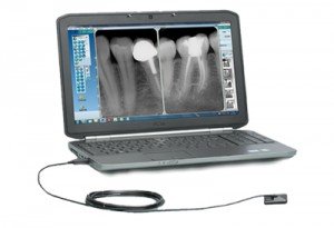
Point x-ray – a maximum of 4 teeth will be visible on this image. This type of x-ray is performed, for example, when the tooth has to have root canal treatment. The photo provides information on the condition and number of root canals and roots, and after the procedure allows to check if the canals have been filled correctly. In addition, the photo provides information on the topic of tooth decay and inflammation.
Panoramic x-ray (OPG) – in the panoramic picture we can see all the teeth and theirs roots, jaw bones, temporomandibular joints and sinuses. OPG allows you to assess the general condition of your teeth, provides information about deep caries, periodontal diseases, retained teeth and broken teeth – in particular wisdom teeth. It also allows you to assess the condition of the bones, which is very important before implant placement.
X-ray in Dental White
At Dental White we use a modern form of teeth X-ray images, from digital radiovisiography (RVG). In this case, the dental x-ray emits up to 10 times lower dose of radiation than traditional shots taken on film, which is much healthier for the patient. Our X-ray is at your servise. You can receive them in the form of outprint or in digital, without additional fees.
RVG advantages
How is RVG performed? The dentist places a special sensor in the patient’s mouth, then a photo is taken, which after a while is visible on the doctor’s computer screen. Pictures taken in RVG technology are very accurate. What’s more, to digitally saved images the doctor has access practically at any time – they are permanently stored on drive. Another important issue is that the doctor can lighten or darken or sharpen the image without any problem, and this makes it easy to read any information.